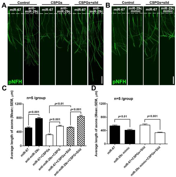
Figure 6.
The effect of miR-29c on axonal growth. Axons in the axonal compartment were transfected with miR-29c inhibitors (A, C) or mimics (B, D). Representative immunofluorescent images (A, B) and quantitative data (C, D) show axonal outgrowth (on DIV5) of distal axons locally transfected with the miR-29c hairpin inhibitor (A, C, anti-miR-29c) or miR-29c mimics (B, D) compared to axons transfected with cel-miR-67 control after axonal application of CSPG and CSPG+sildenafil (CSPGs+sild). Scale bar in A and B=100 μm.

