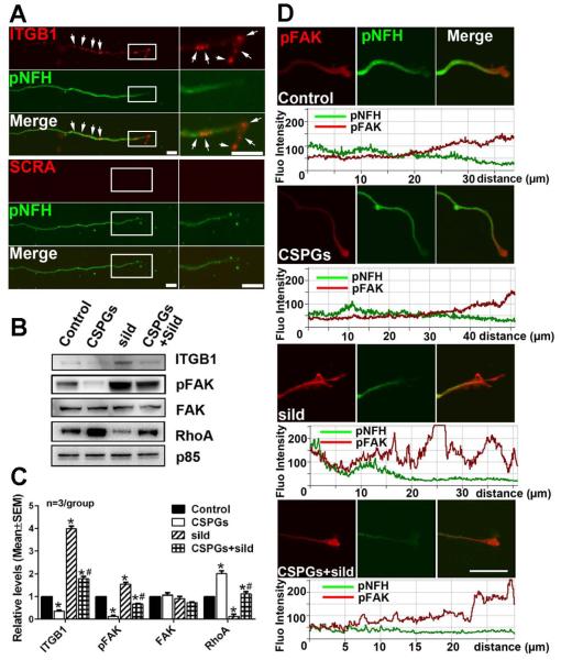
Figure 7.
The effect of axonal application of CSPGs and sildenafil on axonal ITGB1/pFAK/RhoA proteins. Representative FISH in combination with immunofluorescent images (A) show the presence of punctate mRNA signals of ITGB1 (A, red, arrows) in pNFH+ axons (A, green) with low (A, pNFH, green) and high (boxed area in A) magnifications. No signals were detected when FISH was performed with scramble probes (A, SCRA). Representative Western blots (B) and quantification data (C) show levels of ITGB1, pFAK, FAK and RhoA proteins in distal axons after axonal application of CSPGs, sildenafil (sild), or CSPGs+sildenafil (CSPGs+sild). Protein levels of p85 were used as internal controls for the distal axons. * and # p<0.05 vs control and CSPGs, respectively. Representative double immunofluorescent images and their relative fluorescent intensity along with the distal axons (D) show pFAK immunoreactivity (red) of pNFH+ (green) distal axons without any treatment (control) or after axonal application of CSPGs, sildenafil (sild), or CSPGs+sildenafil (CSPGs+sild). The distance (μm) under fluorescent intensity in panel D was measured from the distal axon tip. Scale bar in A=10 μm, in C=20 μm.

