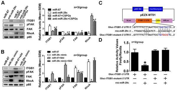
Figure 8.
The effect of miR-29c on on ITGB1/pFAK/RhoA proteins after axonal application of CSPGs or sildenafil in miR-29c reducted axons or miR-29c elevated axons, respectively. Representative Western blots and quantification data show levels of ITGB1, pFAK, FAK and RhoA proteins in distal axons transfected with the miR-29c hairpin inhibitor (A) and miR-29 mimics (B) after the axonal application of CSPGs and sildenafil, respectively. Protein levels of p85 were used as internal controls for the distal axons. * p<0.05 vs control; # p<0.05 vs anti-miR-29c (A) and miR-29c mimic (B), respectively. Panel C shows a schematic of the dual-luciferase reporter vector cloned with segments of the ITGB1 gene 3’UTR. Sequences with blue and red colors are the putative and mutant binding sites, respectively. Panel D shows luciferase activity data from HEK293 cells.* p<0.05 vs Gluc-ITGB1-3’UTR alone transfection group.

