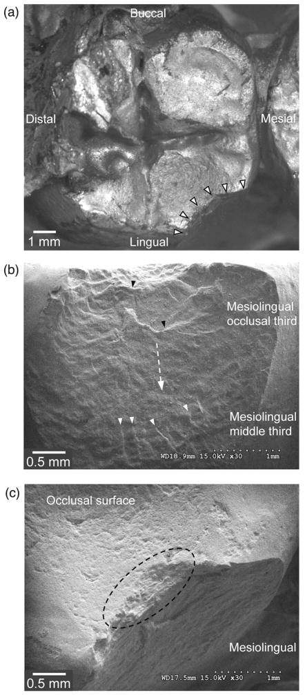
Fig. 1.
A small chipping in the left mandibular second molar (tooth #37, retainer) of a 4-unit zirconia–ceramic FDP. (a) An overview of veneer fracture. Fracture margins are highlighted by white solid arrows. (b) The mesio-lingual view of the fracture surface. The direction of crack propagation is shown by the white dashed arrow. (c) The occlusal view of the fracture origin, highlighted by the dashed circle.

