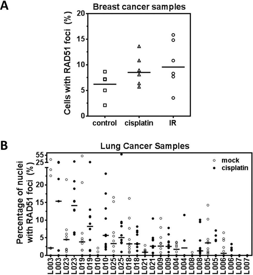
Figure 4.
Penetration of drug into tumor tissue and heterogeneity of RAD51 foci response. A) Example of RAD51 foci responses 5 hours after exposure of breast cancer tissues to cisplatin (8 µM) or ionizing radiation (IR) (10 Gy). Cells with at least two foci were scored as positive. Horizontal lines represent the median (Willers et al., unpublished). B) Analogous to panel A, example of RAD51 foci responses 5 hours after exposure of lung cancer tissues to cisplatin (8 µM). Data points represent foci counts from random high-power images 4.

