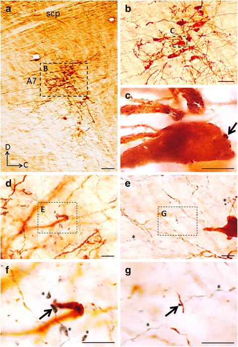
Fig. 2.

BAD-labeled axonal terminals in the A7 area. a. Light microscopic photograph of a rat brainstem section showing BDA deposit in the parabrachial nucleus and TH-ir neurons in the A7. Insert (b) shows a photograph of TH-ir neurons in the A7 with high power. Inserts (c), (e) and (g) show high power photographs of axonal terminals in the A7 area as indicated by dotted squares in the (b), (d) and (f), respectively. Note the terminals of BDA-labeled fibers with prominent en passant type varicosities (asterisks) and the contacts of terminals on large soma in (c), and dendrites in (e, g) as indicated by arrows. d, dorsal; scp, superior cerebellar peduncle. Scale bar = 100 μm (a), 50 μm (b), 10 μm (c-g)
