Fig. 3.

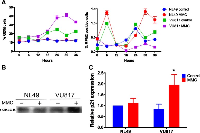
FA cells arrest at G2 phase in response to MMC. a Flow cytometry analysis showing accumulation of FA cells in G2 in response to MMC (left panel) and diminished number of FA mitotic cells compared to normal cells (right panel). b FA cells activate CHK1 kinase in response to MMC treatment. c FA cells increase the expression of p21 mRNA as showed by qRT-PCR analysis (n = 3 independent experiments, p < 0.05)
