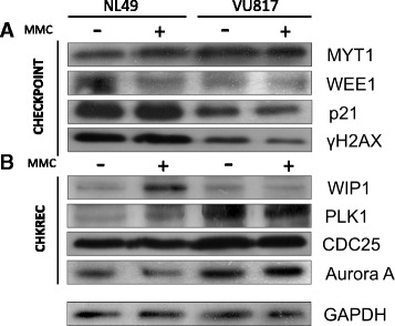
Fig. 6.

FA and normal cells co-express checkpoint and CHKREC proteins. a Western Blot analysis of checkpoint proteins. b Western blot analysis of CHKREC proteins. FA cells increase the amount of some G2 blockage proteins, but have a reduction in others. Although CHK1 (Fig. 4 b) and MYT1 show increased signal, WEE1, γH2AX and p21 protein appear as diminished in FA cells, this weakens the checkpoint blockage, which is eventually overwhelmed by CHKREC signaling (n = 3 independent experiments, see also Fig.7)
