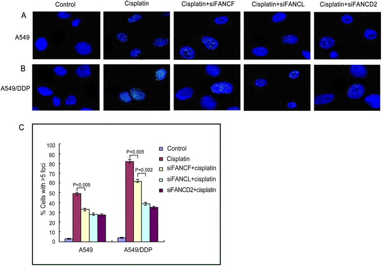
Fig. 2.

Immunofluorescence and quantification of cells exhibiting 5 or more FANCD2 foci in lung cancer cells. Cisplatin-induced FANCD2 foci formation in A549 (a) and A549/DDP cells (b) were inhibited markedly by knockdown of FANCF, FANCL, and FANCD2. c The graph below show quantified data of FANCD2 foci in the two cells. cisplatin-induced FANCD2 foci expressions were stronger in A549/DDP than in A549 cells
