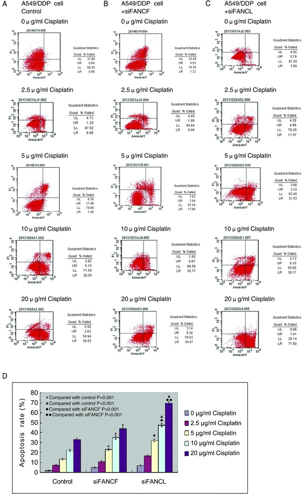
Fig. 7.

Knockdown of FANCF and FANCL promote the apoptosis induced by cisplatin in A549/DDP cells. a The apoptosis rates of control cells, and (b) cells transfected with FANCF-siRNA, and (c) cells transfected with FANCL-siRNA after cisplatin treatment. Early apoptotic cells are in the lower right quadrant; late apoptotic cells are in the upper right quadrant. d The apoptosis rates in cells transfected with FANCF-siRNA or FANCL-siRNA were significantly higher than in control cells in a concentration-dependent manner. In addition, the apoptosis rates of cells transfected with FANCL-siRNA have dramatic increase as compared with cells transfected with FANCF-siRNA following treatment of 10 μg/ml and 20 μg/ml cisplatin
