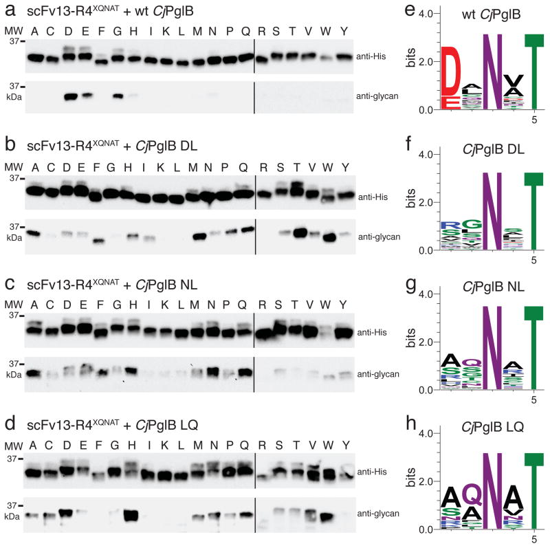
Figure 3. PglB mutants exhibit relaxed substrate specificity.
(a–d) Western blots of acceptor protein scFv13-R4XQNAT, where X is one of the 20 amino acids indicated across the top, co-expressed with each of the CjPglB variants as indicated. The slower migrating band on anti-His immunoblots is the glycosylated form of scFv13-R4XQNAT, confirmed by the anti-glycan immunoblots. Molecular weight (MW) markers are indicated on the left. Blots are representative examples of at least two biological replicates. See Supplementary Fig. 7 for uncropped versions of the images. (e–h) Sequence logos showing experimentally determined substrate specificities of the indicated CjPglB variants from glycoSNAP YebFN24L/XXNXT library screening.

