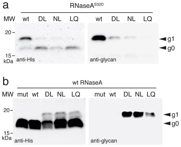
Figure 4. Glycosylation of a native eukaryotic protein by PglB variants.
Western blot analysis of bovine pancreatic RNaseA, with either an S32D substitution in the −2 position of its sequon (a) or its native sequon (b), expressed with each of the CjPglB variants as indicated or with the catalytically inactive CjPglBD54N/E316Q (mut). Molecular weight (MW) markers are indicated on the left. The g0 and g1 labels on the right denote the aglycosylated and glycosylated forms of RNaseA, respectively. Blots are representative examples of at least two biological replicates. See Supplementary Fig. 8 for uncropped versions of the images.

