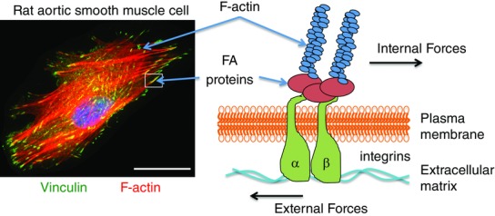
Figure 1.

Vascular smooth muscle cell actin cytoskeleton and adhesion sites
Left-hand panel: immunofluorescence image of a rat aortic smooth muscle cell cultured on collagen-coated glass. Filamentous actin (F-actin) is stained red and vinculin, a focal adhesion (FA) marker, is shown in green. Actin filaments can be seen terminating at adhesion sites. Right-hand panel: schematic diagram of an adhesion site showing sensing of external force and transmission of internal force through actin filaments and integrin focal adhesion site. The scale bar is 0 — 25 μ.
