Figure 1.

Ghrelin inhibits vagal sensory neurons
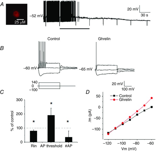
A, continuous membrane potential recording from duodenum-projecting neuron (DiI labelled, inset) showing that ghrelin (30 nm; bar) hyperpolarized the membrane potential and reduced the neuronal input resistance. Negative membrane potential deflections display the neuron potential in response to DC current pulses (−100 pA, 500 ms). B, current responses showing that extracellular application of ghrelin (30 nm) hyperpolarized the recorded neuron. This hyperpolarization was associated with a decrease in neuronal input resistance, as well as a decrease in the number of action potentials evoked by a DC current pulse of amplitude 140 pA (double rheobase) from 6 to 1. C, ghrelin modulates vagal sensory neuron excitability by reducing the neuronal input resistance (Rin), increasing the threshold for action potential initiation and reducing the number of action potentials evoked by a current pulse of the amplitude of double rheobase (n = 8, values are the mean ± SEM, *P < 0.05 compared to controls). D, current–voltage relationships acquired under control conditions and during ghrelin application showing that the effect reversed at approximately −105 mV, close to the estimated K+ equilibrium potential under the conditions recorded.
