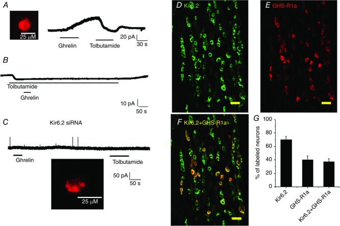
Figure 2.
Ghrelin inhibits vagal sensory neurons via activation of KATP channel
A, continuous current recording showing that tolbutamide (200 μm), a KATP channel inhibitor, reversed the current induced by ghrelin (30 nm) in a duodenum-projecting neuron (DiI labelled, inset). B, representative continuous current recording showing that extracellular application of tolbutamide (200 μm) generated an inward current (10 pA). Subsequent application of ghrelin (30 nm) failed to induce an outward current. C, silencing the Kir6.2 channel subunit in a vagal sensory neuron using Kir6.2 Cy3-labelled siRNA abolished both ghrelin and tolbutamide currents. The inset shows a Kir6.2 Cy3-siRNA transfected/labelled neuron. D–F, photomicrographs showing anti-Kir6.2 (green) and anti-GHS-R1a (red) immunoreactivities in vagal ganglia cross-sections. Superimposition of the green and red images showing that most anti-GHS-R1a immunoreactivity colocalizes with anti-Kir6.2 (yellow). Scale bar, 100 μm. G, percentage of neurons in vagal ganglia expressing Kir6.2 and GHS-R1a, as well as both Kir6.2 and GHS-R1a. The data are based on counts from five cross-sections.

