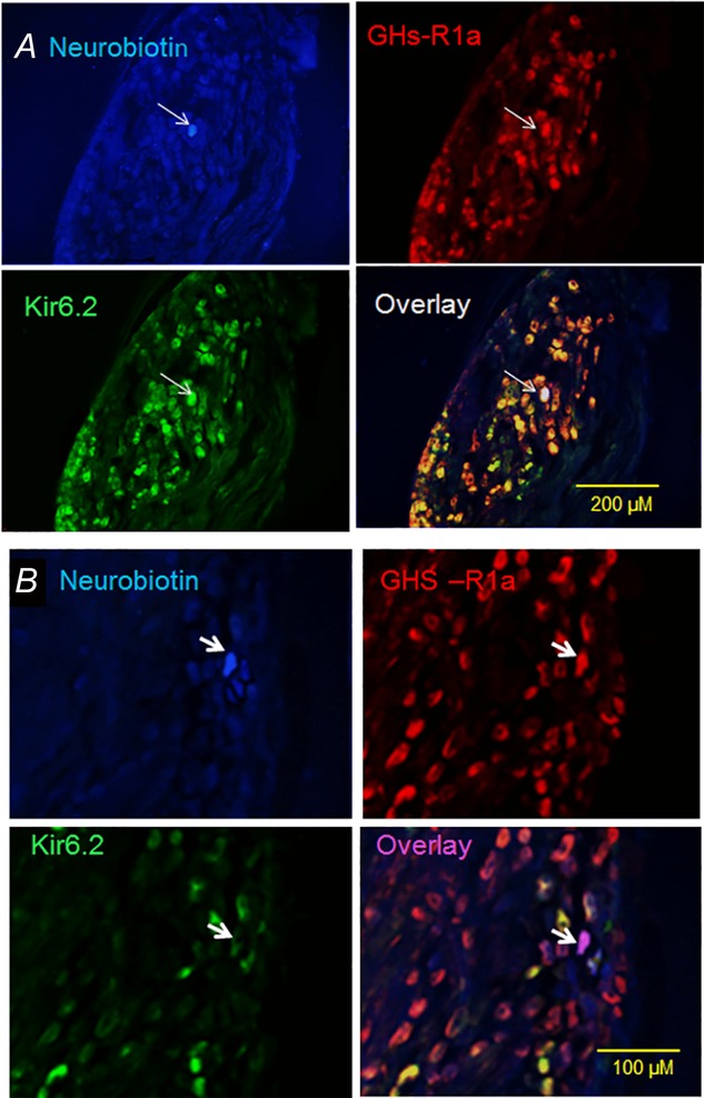
Figure 7.

Immunostaining of rat nodose ganglia showing neurons targeted for electrophysiological recording
A, photomicrographs of vagal ganglia cross-sections showing a neuron (arrow) injected with neurobiotin (blue) after electrophysiological recording. This neuron expresses both anti-Kir6.2 (green) and anti-GHS-R1a (red) immunoreactivities. Superimposition of the green and red images (overlay) showing that most of the anti-GHS-R1a immunoreactivity colocalizes with anti-Kir6.2 (yellow). Note that the recorded neuron labelled with neurobiotin contains Kir6.2 and GHS-R1a immunoreactivities (white). B, photomicrographs showing a neuron (arrow) injected with neurobiotin (blue) after electrophysiological recording in a Kir6.2 siRNA-electroporated animal. This neuron exhibits strong immunoreactivity to anti-GHS-R1a (red) but little immunoreactivity to anti-Kir6.2 (green). Note that few neurons in the overlay image coexpress Kir6.2 and GHS-R1a (yellow). The recorded neuron (arrow in overlay image) shows no immunoreactivity to Kir6.2 but expresses GHS-R1a (purple).
