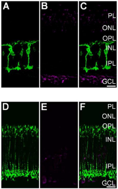
Figure 5.

Light-evoked S-nitrosylation in the goldfish and wild-type mouse retina is prevented by pre-incubation with N-Ethylmaleimide (NEM). A: Single plane 40× confocal image showing PKCα + (green) Mbs in a vertical cryosection from a goldfish retina incubated in 1 mM NEM for 20 min prior to mesopic light stimulation (1×10¹⁰ photons/cm2/s, 505 nm, 10 sec). B: Same region as in A showing S-nitrosocysteine immunolabeling (magenta) in response to mesopic light stimulation. Note absence of immunolabel signal when compared to the untreated goldfish retina seen in Fig. 3A, B, and C. C: Merged image of confocal images from A and B showing very little colocalization between PKCα + Mbs and S-nitrosocysteine immunofluoresence. D: Single plane 40× confocal image showing PKCα + (green) RBCs in a vertical cryosection from a wild-type mouse retina incubated in 1 mM NEM for 20 min prior to mesopic light stimulation (1×10¹⁰ photons/cm2/s, 505 nm, 10 sec). E: Confocal image of the same region as presented in A illustrating that the mesopic light-evoked SNI (magenta) is dramatically reduced as compared to the untreated wild-type mouse retina (Fig. 4A, B, and C). F: Merged 40× confocal imaged of a wild-type mouse retina co-immunolabeled for PKCα and S-nitrosocysteine. Scale bars=20 μm.
