
Figure 10.
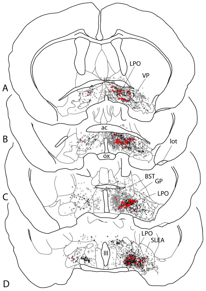
Map showing several sections through the basal forebrain ordered rostrocaudally from A to D and illustrating retrogradely labeled neurons following injection of the retrograde tracers cholera toxin β subunit into the rostromedial tegmental nucleus (circles) and Fluorogold into the lateral habenula (crosses). Neurons exhibiting both tracers (double-labeled) are shown as red dots. Note that the double-labeled neurons are located preferentially in the lateral preoptic area, which probably reflects the greater concentration of retrogradely labeled neurons in the lateral preoptic area following lateral habenula injections of tracer (see also Fig. 11 and Table 6). See list for additional abbreviations.