
Figure 5.
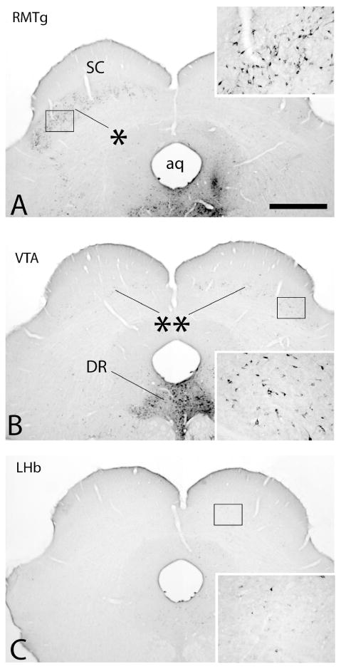
Photomicrographs illustrating retrograde labeling in the deep layers of the superior colliculus (SC) following injections of cholera toxin β subunit (Ctβ) into the rostromedial tegmental nucleus (RMTg in A), ventral tegmental area (VTA in B) and lateral habenula (LHb in C). Insets are enlargements of the boxed areas in the respective micrographs. Tracer was injected into the brain to the right of the midline. Note following injection of tracer into the RMTg that retrograde labeling is distributed almost exclusively contralateral to the RMTg injection (asterisk in A). Substantially less dense labeling was distributed bilaterally but with an ipsilateral bias following tracer injection into the VTA (double asterisks in B). Following LHb injections, labeling in the SC was almost entirely ipsilateral and, although there were moderate numbers of labeled neurons, they were very sparsely labeled with immunoperoxidase reaction product, such that higher magnification was necessary to visualize them (inset in C). See list for additional abbreviations. Scale bar: 1 mm.