
Figure 8.
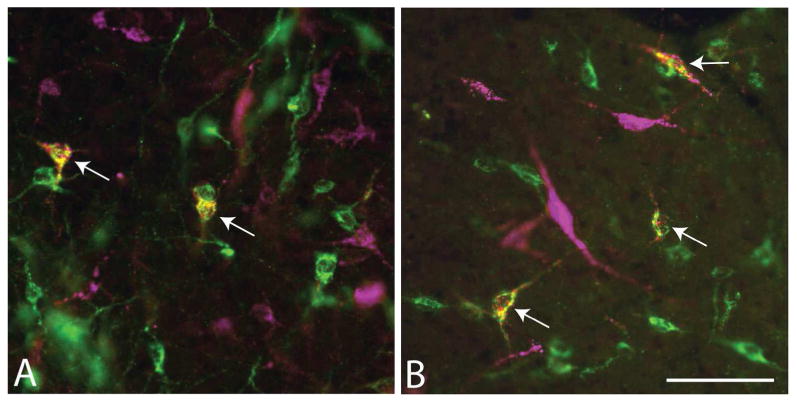
Micrographs showing single-labeled neurons (green and magenta) and double-labeled neurons (white arrows) in the lateral preoptic region following injections of (A) cholera toxin β subunit (Ctβ) into the VTA and Fluorogold (FG) into the RMTg (map is shown in Figure 9) and (B) FG into the LHb and Ctβ into the RMTg (map is shown in Figure 10). Scale bar: 100 μm.