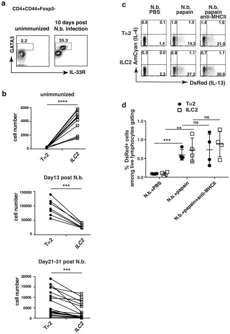
Figure 4. TH2 cells generated by infection with N. brasiliensis respond to papain to produce TCR-independent IL-13.
(a) Lung TH2 cells expanded dramatically upon N. brasiliensis infection. Lungs were harvested from mice either unimmunized or 10 days after N. brasiliensis infection. TH2 cells were identified as CD4+CD44+Foxp3−GATA3+IL-33R+. Markers for ILC2 cells were Lin−CD44+GATA3+IL-33R+CD127+Thy1+. Data are representative of at least three independent experiments with 3 mice in each group. (b) Number of lung-resident TH2 and ILC2 cells in individual mice that were unimmunized or 13 days or 21–31 days post-N. brasiliensis infection. ****, P<0.0001; ***, P<0.001 by paired t-test. Data are compiled from multiple independent experiments. (c, d) Twenty-five days after N. brasiliensis infection, 4C13R reporter mice were challenged intratracheally with PBS, or papain (25 μg in PBS) for 3 consecutive days with or without anti-MHCII antibody. 500 μg anti-MHCII antibody was administered i.v. on day 1 and day 3 of papain challenge. DsRed and AmCyan expression by lung-resident TH2 and ILC2 cells were analyzed 24h after last papain challenge. (d) Comparison of percent of DsRed+ TH2 and ILC2 cells among total live lymphocytes. ns, not significant, P>0.05; **, P<0.01; ***, P<0.001. Data are representative of three independent experiments with 4 mice in each group (c–d).

