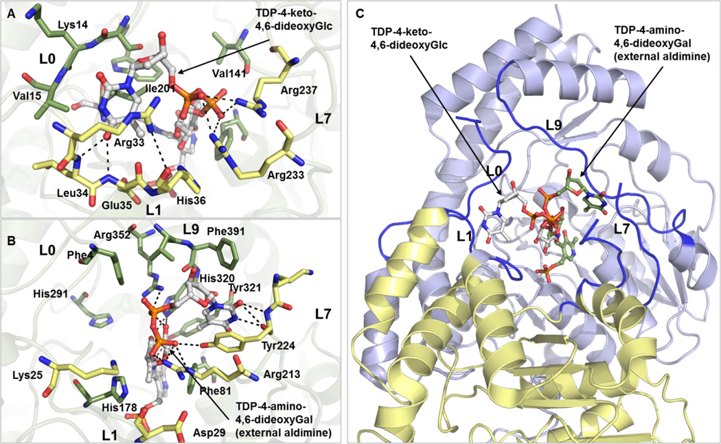
Figure 5.
Nucleotide sugar binding site of (A) CalS13 and (B) WecE. The substrates within the CalS13 and WecE active sites are represented as ball and stick models and colored white. The active site residues from the same and adjacent subunits are colored green and yellow, respectively. The spatial locations of loops L0, L1, L7, and L9 are labeled. (C) Overlay of the orientation of binding of TDP-4-keto-4,6-dideoxy-α-d-glucose from CalS13 and TDP-4-amino-4,6-dideoxy-α-d-galactose as external aldimine from WecE in the active site of CalS13. The TDP-4-keto-6-deoxy-α-d-glucose and the external aldimine form of TDP-4-amino-4,6-dideoxy-α-d-galactose are represented as ball and stick models colored white and green, respectively. The loops L0, L1, L7, and L9 are colored blue, and the two SAT subunits are colored blue and yellow.

