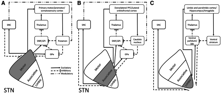
Fig. 1.
Schematic illustration of positioning of the STN and the connectivity of functional putative subdivisions within the basal ganglia thalamocortical a motor, b associative, and c limbic circuits. Adapted from (Temel et al. 2005). Based on tracing studies in monkeys using both antero- and retrograde tracings, anatomofunctional subdivisions of the STN have been proposed. Cortical areas involved in the motor circuitry include the primary motor, premotor, and somatosensory cortex (a). Cortical areas involved in the associative circuitry include the dorsolateral prefrontal cortex, as well as the lateral orbitofrontal cortex (b). The associative circuits include the direct and indirect pathway. The direct pathway runs via the internal segment of the Globus Pallidus (GPi) and reticular part of the Substantia Nigra (SNr) to the ventroanterior (VA) and centromedian (CM) nuclei of the thalamus, and the circuit is closed by the thalamocortical pathway back to the dorsolateral prefrontal cortex (DLPC) and the lateral orbitofrontal circuit back to the lateral orbitofrontal cortex (LOFC). The indirect pathway encompasses a projection from the external part of the globus pallidus (GPe) to the STN and GPi/SNr. The limbic circuitry involves limbic and paralimbic cortices as well as hippocampus and amygdala (c) (Temel et al. 2005). Although subdivisions are anatomically separated in this illustration, evidence from tracing studies point towards significant overlap of these subdivisions (Haynes and Haber 2013)

