Fig. 7.
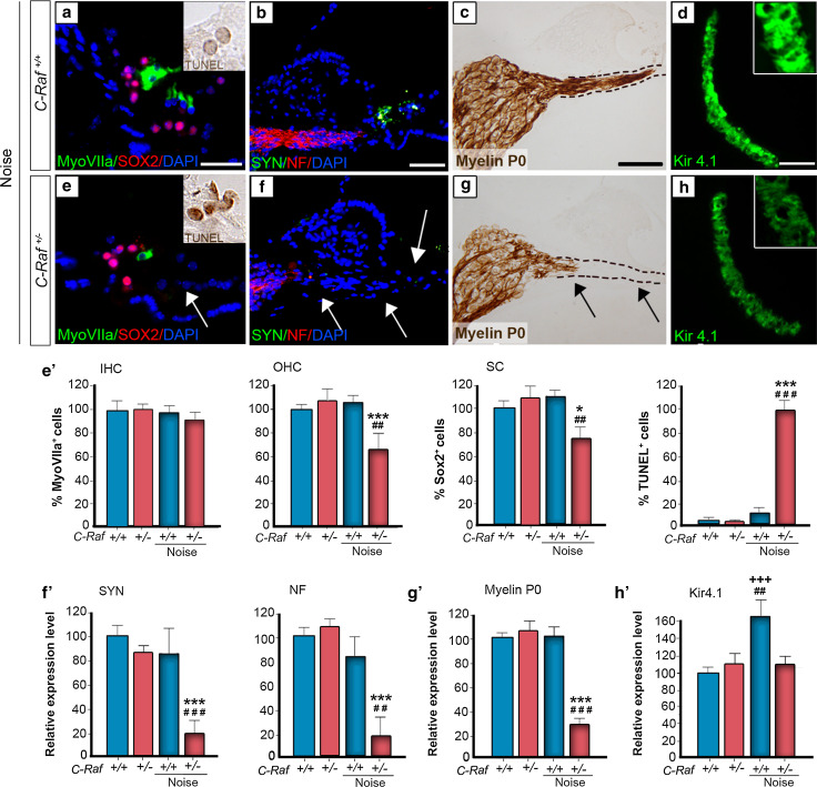
C-Raf heterozygous mice exhibited severe cellular alterations in the organs of Corti and spiral ganglion following noise exposure. a, e, e′ Wild-type and heterozygous mice exposed to noise showed different immunostaining patterns. Myosin VIIa (green) positive outer hair cells and SOX2 (red) positive supporting cells showed 40 and 25 % reductions, respectively, in heterozygous mice. In contrast, TUNEL staining 2 days after noise exposure (insets a, e) showed a 95 % increment in the noise-exposed heterozygous mice with respect to any other experimental group (e′). b, c, f, g, f′, g′ Neuronal markers NF (red), SYN (green) and myelin P0 (brown) evidenced neuronal and nerve fiber loss. Quantification of staining/μm2 showed that only heterozygous mice exposed to noise presented a general reduction. d, h, h′ Immunostaining for the Kir4.1 potassium channel (green) in the stria intermediate cells increased by 60 % in the wild-type but not in the heterozygous mice after noise exposure. The intensity of the immunofluorescence per μm2 of the regions was quantified using ImageJ software. Data were obtained from 4 to 12 sections of at least three mice from each genotype and are shown relative to those of non-exposed wild-type mice as mean ± SEM. The significance of the differences was evaluated using Student’s t test: +++ P < 0.001 versus non-treated wild type; *P < 0.05 versus non-treated heterozygous; ***P < 0.001 versus non-treated heterozygous; ## P < 0.01 versus wild type exposed to noise; ### P < 0.001 versus wild type exposed to noise; Scale bars 10 µm (a, e); 25 µm (b–d, f–h)

