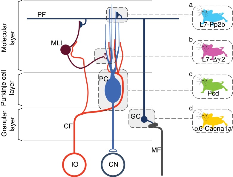
Fig. 1.
Microcircuitry of the cerebellar cortex highlighting the main sites affected in the Pcd, L7-Pp2b, L7-∆γ2 and α6-Cacna1a mice. The two main excitatory afferents of the cerebellar cortex are the mossy fibers (MF) and climbing fibers (CF). Whereas the MFs originate from various sources in the brainstem, all CFs are derived from the inferior olive (IO). The CFs directly innervate the Purkinje cells (PCs) and influence via non-synaptic release the activity of molecular layer interneurons (MLI), which inhibit PCs. The MFs directly innervate the granule cells (GCs), which in turn give rise to the parallel fibers (PFs) that innervate both PCs and MLIs. PCs form the sole output of the cerebellar cortex to the cerebellar nuclei (CN). The mutants used in the current study either lack Purkinje cells (Pcd, indicated in green), intrinsic Purkinje cell plasticity and parallel fiber-to-Purkinje cell potentiation (L7-Pp2b, blue), phasic inhibition provided by molecular layer interneurons (L7-∆γ2, purple), or most of their granule cell output (α6-Cacna1a, yellow)

