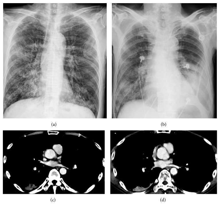
Figure 3.
Pulmonary vein obstructive syndrome (PVOS). A 51-year-old man with oral cancer. Chest X-ray (a) before and (b) at the onset of PVOS showed a left low lung and hilum increase haziness. CT scan ((c) and (d)) revealed tumor/thrombosis located in superior and inferior pulmonary veins with left atrium extension.

