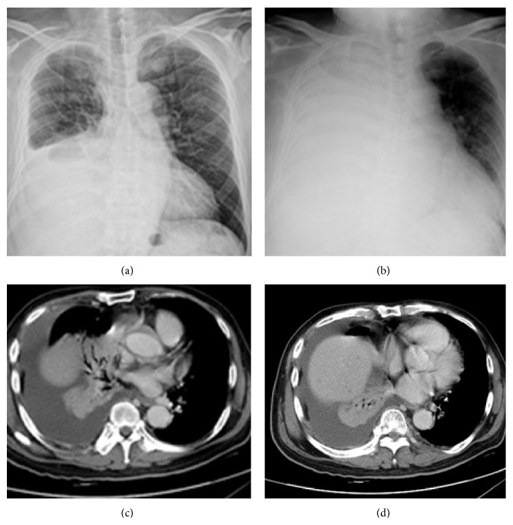
Figure 4.
Pulmonary vein obstructive syndrome (PVOS). A 61-year-old man with lung cancer. Chest X-ray (a) before and (b) at the onset of PVOS showed right low lung increase haziness to total opacity. CT scan ((c) and (d)) revealed lung tumor and atelectatic lesions surrounding the superior and inferior pulmonary veins with right pleural effusion.

