Figure 1.
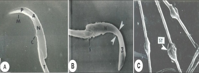
Scanning electron micrographs (SEM) of cauda epididymal spermatozoa of control (Fig.A) and treated with 250mg/kg body weight of benzene extract rat (Figs. B&C)
A. Spermatozoa of control rat exhibiting normal parts of acrosome (A); post nuclear cap (C); plasma membrane (M); nucleus (N) perforatium (P) and tail region (T). 4.56kx.
B. The perforatium (sub-acrosomal material), swells (PS) and the middle region of the sperm head is constricted dorsoventrally (arrows). There is serration (S) at the connective piece of the spermatozoa. A5.56kx.
C. There is increase of swelling at tail region in the mid portion of the tail region rat spermatozoa (arrows, ST). 5.93 kx.

