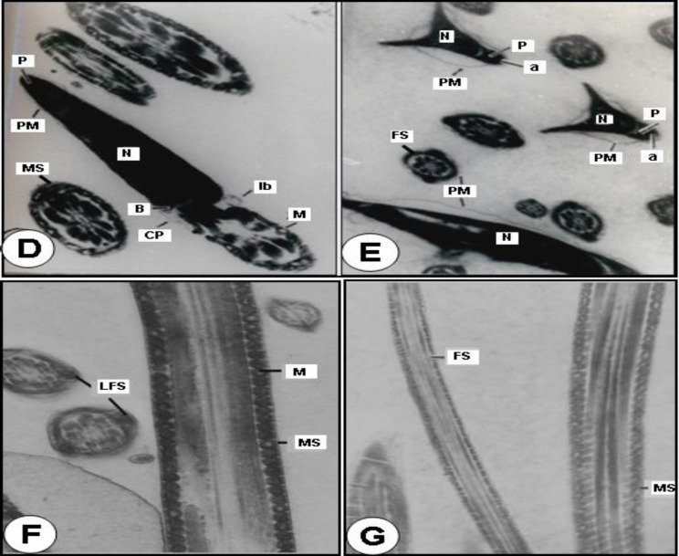
Figure 2.
Transmission electron micrographs (TEM) of the control (D-G).
D: A median section through the base of the sperm head illustrating nucleus (N) covered with perforatorium (P), basal plate (B), plasma membrane (PM) and connecting piece of tail (CP). Caudal portion of the nucleus illustrating lamellar body (lb) and mitochondria (M) are seen, in oblique section, in contact with connecting piece. Cross sections(CS) of mid-piece of spermatozoa flagellum with spatial arrangement of the 9 outer dense fibres to one another, to the centrally located axoneme composed of the 9 plus 2 arrangements of microtubules and to the mitochondrial sheath (MS) X 29,000.
E. C.S. of anterior portion of sperm head illustrate normal features of nucleus (N); perforatorium (P); acrosome (a); plasma membrane (PM) and principal piece are with normal features of fibrous sheath (FS) and arrangement of outer fibres and axonemal component X 21,700.
F-G: Longitudinal section (LS) of mid piece and principal piece illustrating well-preserved mitochondrial sheath (MS) and fibrous sheath (FS) and are intact with plasma membrane. Mitochondria (M) appear normal. C.S. of principal piece is with normal features of longitudinal fibrous sheath (LFS) and arrangement of outer fibres and axonemal component X 15,000.

