Figure 3.
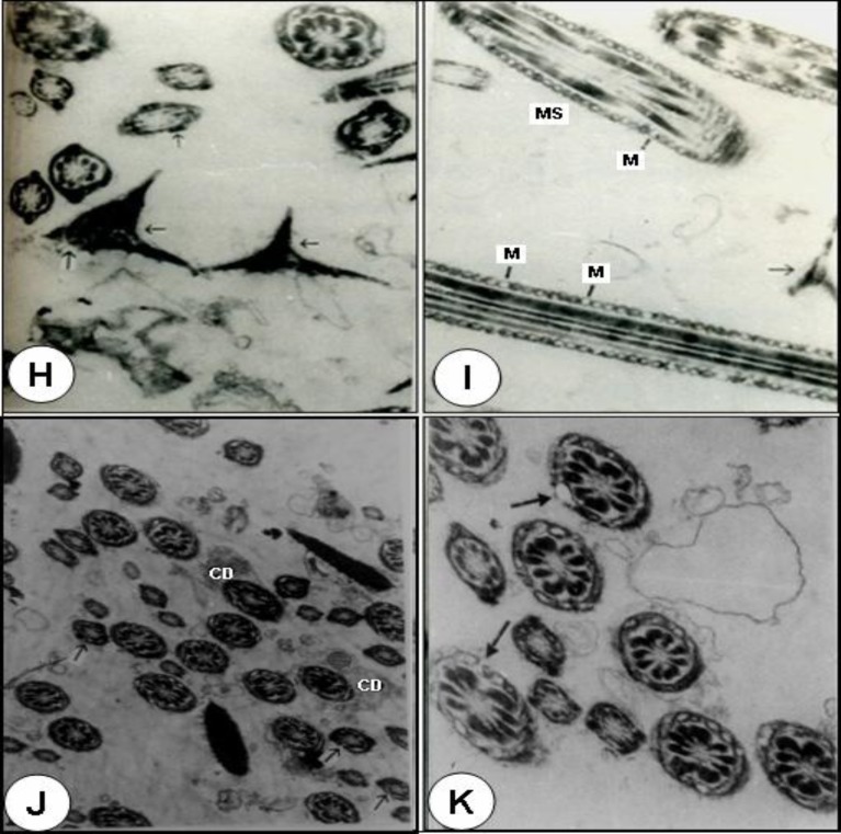
Treated with 250mg/kg body weight of benzene extract rat (Figs. H-K).
H: C.S. of the different parts of sperm head (←) showing disruption in normal features plasma membrane, acrosome and perforatorium. Fuzzy material is seen on their surface. X 15,000.
I: L.S and C.S. of the mid piece shows the loss of plasma membrane. Most of mitochondria (M) are hypertrophied or have started degeneration. The loss of plasma membrane is seen in the sperm head region (←) X 28,000.
J: C.S. of the mid piece showing abnormal pattern of mitochondrial sheath and loss of plasma membrane. Most of mid-piece show increased cytoplasmic droplets (CD) displaced on one side are clearly seen. Coating of fuzzy material is seen on the surface of head and tail sections X 10,300.
K: C.S of the mid piece shows abnormal pattern and degeneration of mitochondrial sheath (long arrows) along the length of the structure and displaced mitochondrial sheath on one side. The discontinuation of fibrous sheath is also noticed in principal piece X 17,500.

