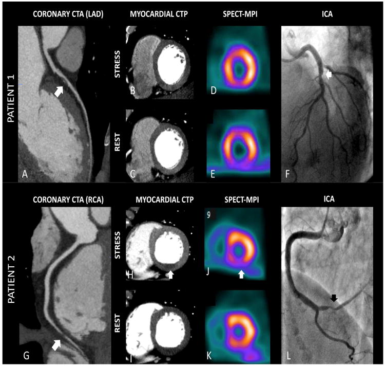
Figure 2.

Combining CTA and CTP data in diagnostic of flow-limiting stenosis. Patient 1 - (A) Left Anterior Descending (LAD) artery with a mixed plaque leading to an intermediate (50-69%) stenosis (CTA positive for ≥50% stenosis - arrow). (B, C) Stress/Rest images showing no evidence of perfusional deficits in anterior wall, with correspondent findings in SPECT-MPI (combined CTA + CTP negative for ≥50% stenosis with related perfusion deficit) (D, E). (F) Invasive angiography showing borderline stenosis in LAD (arrow), but with no relation to myocardial ischemia according to SPECT results (reference standard negative for ≥50% stenosis with related perfusion deficit). Patient 2 – (G) Significant stenosis in distal Right Coronary Artery (RCA) (CTA positive for ≥50% stenosis - arrow), with a corresponding perfusional deficit (most reversible) in left ventricle inferior wall ((H, I). Combined CTA and CTP was positive for ≥50% stenosis with related perfusion deficit. Reference standard images show similar reversible perfusion deficit in inferior wall by SPECT-MPI (J, K), and the related stenosis by ICA (L). CTA - Computed Tomography Angiography; CTP – Computed Tomography Perfusion; ICA – Invasive Coronary Angiography; SPECT –MPI – Single-Photon Emission Computed Tomography – Myocardial Perfusion Imaging.
