
Figure 6. Consistency of theta generation across cortical locations.
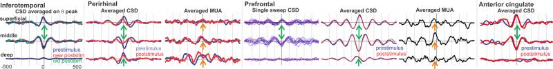
CSD, averaged on the peak of the theta rhythm, shows that in all 4 cortical areas, the main currents generating the theta are a middle layer sink paired with a superficial source and a weaker deep source. Correlated increases in MUA (in two areas) imply that the sink is active excitation. Spontaneous (prestimulus) and evoked (poststimulus) theta distributions are identical, demonstrating that their neural generators are the same at a microphysiological level. Recordings from each cortical area was obtained in a different 4 subject.