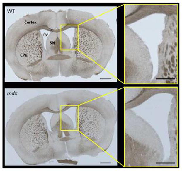
Figure 3.
Representative sections of brain from WT and mdx mice. Gross morphology was similar in both groups (N = 2 per group), but the changes in ventricle size seen with imaging (Fig. 2) were also apparent with histological imaging. CC = corpus callosum, CPu = caudate putamen, RV = right ventricle, SN = septal nucleus. Image scale bars = 1000 μm. Panel inset scale bars = 500 μm.

