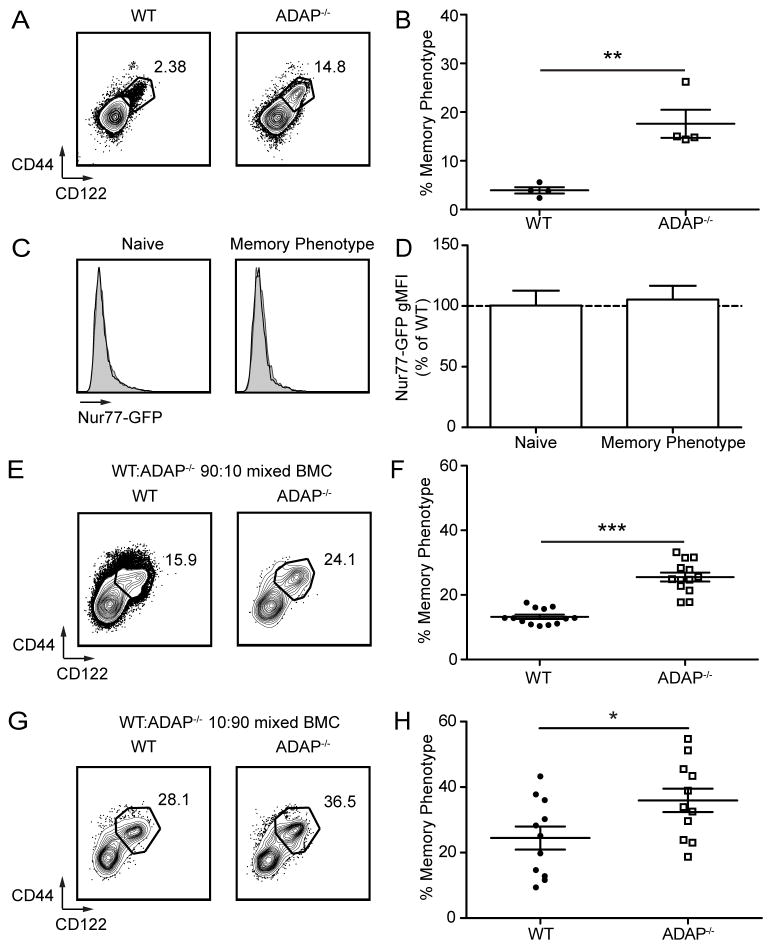
Figure 4. Memory phenotype in the absence of ADAP is not due to CD8 T cell self-reactivity, and is specific to ADAP-deficient CD8 T cells.
(A) CD44 and CD122 staining of spleens from WT and ADAP−/− Rag2−/− P14-Tg mice. Numbers represent the percentage of MP cells. Analyzed cells are CD8+ CD4− Vα2+ cells. (B) Percentage of MP WT and ADAP−/− Rag2−/− P14-Tg T cells. (C) Spleens from littermate polyclonal WT and ADAP−/− mice expressing the Nur77-GFP reporter were harvested and the expression of Nur77-GFP was analyzed by flow cytometry in CD8+CD44lo/CD122lo (Naïve) and CD8+CD44hi/CD122hi (MP) cells. Representative histograms depict Nur77-GFP expression for WT (black line) overlaid with ADAP−/− (grey shaded). (D) Quantification of Nur77-GFP fluorescence of naïve and MP CD8+ T cells from 4 separate littermate pairs of WT and ADAP−/− mice analyzed in two independent experiments. Values are normalized to Nur77-GFP expression observed in WT mice. (E–H) WT (CD45.1/2) and ADAP−/− (CD45.2) bone marrow cells at the indicated ratios were transferred into lethally irradiated CD45.1 recipients. Mixed bone marrow chimeras were harvested 8–12 weeks after transfer of donor marrow. (E–F) WT: ADAP−/−, 90:10 ratio, (G–H) WT: ADAP−/−, 10:90 ratio. (E and G) CD44 and CD122 staining of CD8+ CD4− TCR-β + donor cells from recipient spleens. Numbers represent the percentage of MP cells. (F and H) Percentage of CD44hi CD122hi donor cells. The results (B) are compiled from three independent experiments (± SEM) with at least two mice per experiment, (D) is compiled from three independent experiments, each with at least one littermate pair, and (F and G) are compiled from three independent experiments, each with at least 4 mice.

