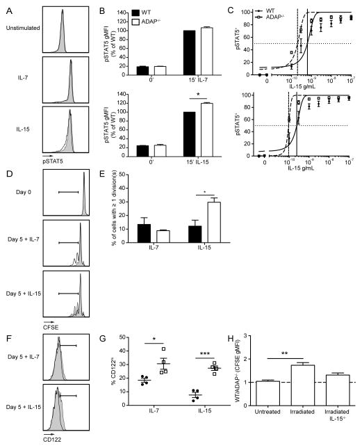
Figure 8. Mature naïve CD8 T cells are more responsive to IL-15 in the absence of ADAP.
(A–C) Bulk splenocytes were obtained from WT and ADAP−/− adult mice and stained for intracellular pSTAT5. (A) Histograms of WT (black) or ADAP−/− (grey shaded) pSTAT5 staining on CD8+ CD122lo cells after addition of 1 ng/mL of IL-7 or IL-15 for 15 minutes. (B) STAT5 phosphorylation was measured by flow cytometry and quantified as the population gMFI staining normalized to WT IL-7 or IL-15 stimulated samples from CD8+ CD122lo cells. (C) Dose response to IL-15 (0.1–300 ng/mL) for 15 minutes in CD8+ CD122lo cells (top) and CD8+ CD122hi cells (bottom) LogEC50 for CD8+ CD122lo cells was 6.3 × 10⁻¹⁰ (WT) and 2.0 × 10⁻¹⁰ (ADAP−/−), For CD8+ CD122hi cells, the LogEC50 was 2.4 × 10⁻¹⁰ (WT) and 9.4 × 10⁻¹¹ (ADAP−/−). (D–G) Naïve CD8 T cells were isolated from WT (CD45.1/2) or ADAP−/− (CD45.2) OT-I adult mice, mixed at a 1:1 ratio, and labeled with CFSE. WT and ADAP−/− cells were co-cultured in the presence of 10 ng/mL of IL-7 or IL-15 for 5 days. (D) CFSE staining of WT and ADAP−/− CD8 T cells at day 0 or 5 after addition of cytokine. Gate indicates cells that have undergone 1 or more divisions. (E) Percentage of donor cells that have undergone one or more divisions. (F) CD122 staining of WT and ADAP−/− CD8 T cells at day 5 after addition of cytokine. Gate indicates cells that are CD122hi. (G) Percentage of CD8 T cells with expression of CD122hi. (H) Naïve OT-I CD8 T cells from pLNs were isolated from WT (CD45.1) and ADAP−/− (CD45.1/2) adult mice, labeled with CFSE, and co-transferred into untreated or sub-lethally irradiated CD45.2 or IL-15−/− CD45.2 recipients. Recipient spleens were harvested 7 days after transfer. Ratio of WT:ADAP−/− CFSE gMFI in untreated or sub-lethally irradiated wild-type or IL-15−/− recipient spleens. The results (B, C, E, G and H) are compiled from three independent experiments (± SEM).

