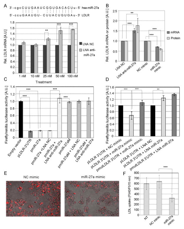
Fig. 4. Effect of miR-27a on the expression of LDLR in HepG2 cells.
(A) Predicted annealing of human miR-27a to two sites on the LDLR 3′UTR. HepG2 cells were transfected with different concentrations of either LNA anti-miR-27a or LNA NC. (B) Cells were transfected with either 30 nM miR-27a, 30 nM NC mimic, 50 nM LNA anti-miR-27a or 50 nM LNA NC. The level of LDLR mRNA was quantified by TaqMan qPCR and the LDLR protein by ELISA. (C and D) Cells were co-transfected with 1 μg of reporter constructs pLDLR-3′UTR, pmiR-27a or mutated pmiR-27aM in the presence or absence of either 50 nM LNA anti-miR-27a, 50 nM LNA NC, 30 nM miR-27a or 30 nM NC mimic. (E) LDLR activity was assessed in HepG2 cells transfected with either 30 nM miR-27a or NC mimics and incubated overnight with LDL conjugated to DyLight™ 549, a fluorescent probe for detection of LDL uptake. The degree of LDL uptake was examined in an Evos Digital Inverted Fluorescence Microscope (AMG, Fisher Scientific) with filters for excitation at 540 nm and emission at 570 nm. (F) The intensity of fluorescence was quantified at 540/570 nm excitation/emission using a Synergy MX plate reader (Biotek). Data are mean of three independent experiments ± S.D. LNA, locked nucleic acids; NC, negative control; LDLR, LDL receptor. The significance is indicated only for samples that are significantly different from all the others. * P<.05; ** P<0.01; *** P<0.001.

