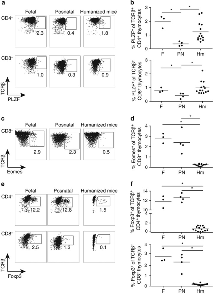
Figure 2.
Non-conventional T-cell development in humanized mice is distinct from fetal and postnatal human thymopoiesis. (a) Representative flow-cytometric analysis of PLZF expression in CD4+ (top panels) and CD8+ (bottom panels) human thymocytes gated on TCRβ+ cells in fetal, postnatal and humanized mice thymic samples. (b) Proportion of PLZF+ cells in CD4+ (top panel) and CD8+ (bottom panel) TCRβ+ human thymocytes in human fetal, postnatal and humanized mice thymic samples. Each dot represents one thymus. (c) Representative flow-cytometric analysis of Eomes expression in CD8+ thymocytes gated on human TCRβ+ cells in human fetal, postnatal and humanized mice thymic samples. (d) Proportion of Eomes+ cells in CD8+ TCRβ+ human thymocytes in human fetal, postnatal and humanized mice thymic samples. (e) Representative flow-cytometric analysis of Foxp3 expression in CD4+ (top panels) and CD8+ (bottom panels) human thymocytes gated on TCRβ+ cells in fetal, postnatal and humanized mice thymic samples. (f) Proportion of Foxp3+ cells in CD4+ (top panel) and CD8+ (bottom panel) TCRβ+ human thymocytes in human fetal, postnatal and humanized mice thymic samples. Line represents average. * indicates P<0.05.

