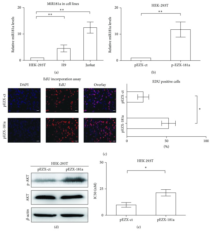
Figure 3.
Ectopic expression of miR181a enhanced cell proliferation and resistance to doxorubicin through AKT activation. (a) T-leukemia/lymphoma Jurkat and H9 cells had significantly higher expression levels of miR181a than that of HEK-293T cells. ** P < 0.01 comparing with HEK-293T cells. (b) Transfection with miR181a (pEZX-181a) in HEK-293T cells resulted in significantly increased miR181a expression. ** P < 0.01 comparing with the control pEZX-ct cells. (c) EdU incorporation assay in HEK-293T cells showed that miR181a-overexpressing pEZX-181a cells presented with increased EdU-positive cells. * P < 0.05, comparing with the control pEZX-ct cells. Bar = 20 μm. (d) Overexpression of miR181a increased AKT phosphorylation, while the total protein level remained constant. (e) IC50 of doxorubicin was significantly higher in the pEZX-181a cells than in the control pEZX-ct cells. * P < 0.05 comparing with the control pEZX-ct cells.

