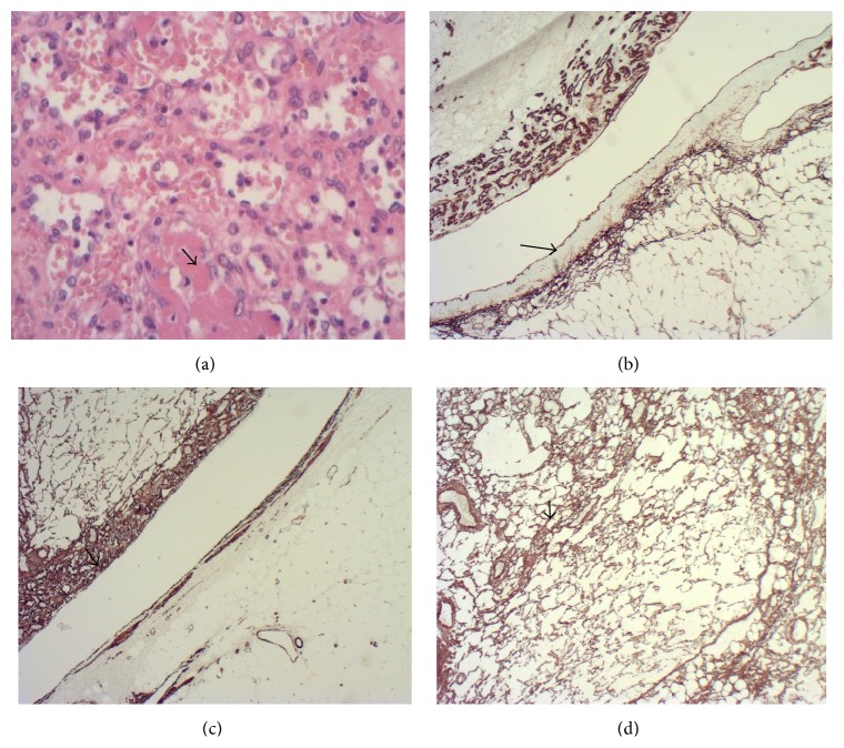
Figure 3.
(a) 200x H&E showing fibrin thrombi within some vascular spaces. (b) showing CD34 immunohistochemistry staining the endothelium in the tumour as well as the segmental renal vein branch (arrowed) (×20). (c) demonstrating smooth muscle actin immunohistochemistry staining the tumour pericytes (arrowed) and vessel wall (×20). (d) showing CD34 immunohistochemistry staining supporting pericytes (×40).

