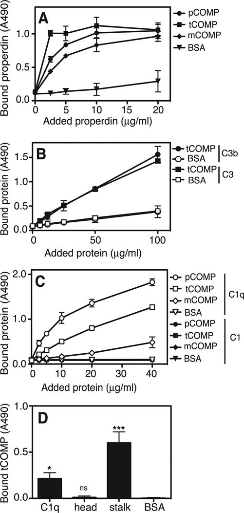
FIGURE 3. COMP binds to properdin, C3 and C1q.
tCOMP, pCOMP and mCOMP were coated onto microtiter plates and incubated with fluid phase properdin at increasing concentrations (A). To compare binding of C3 and the C3b activation product to tCOMP, tCOMP was immobilized and increasing concentrations of C3 and C3b were added to the plate. BSA was immobilized as a negative control (B). In order to compare binding of COMP to C1 and C1q, tCOMP, pCOMP and mCOMP were coated onto microtiter plates and fluid phase C1q or C1 at increasing concentrations was allowed to attach (C). To examine the binding region for COMP on C1q, isolated head and stalk fragments of C1q or intact C1q were immobilized and incubated with 20 µg/ml tCOMP (D). The data are given as the mean and SD of three separate experiments. Statistical significance was calculated using a one-way ANOVA in panel D. ns, not significant, *, p < 0.05, **, p < 0.01, ***, p < 0.001.

