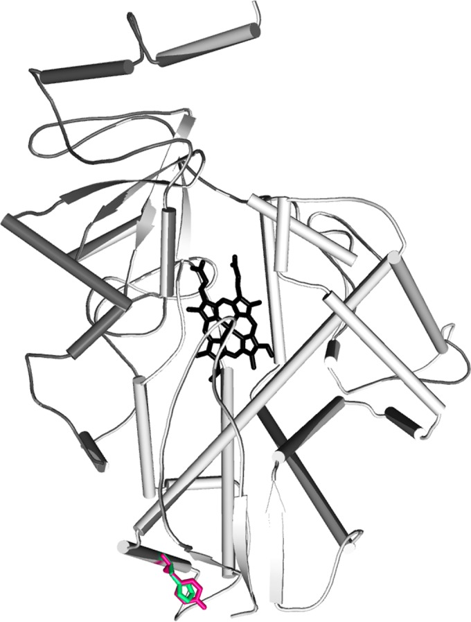
FIG 1.

Modeled structure of CYP51C of A. flavus shown in cartoon representation. The porphyrin ring is shown in stick representation in black. The tyrosine residue present in the wild type and the histidine in the mutant are shown in hot pink and green, respectively.
