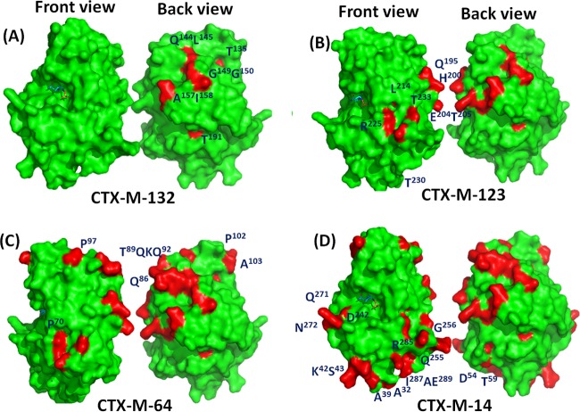
FIG 5.
Structural models depicting the locations of divergent residues. Front and back views of the proteins are shown. The front view shows the active site side of protein, while the back view is distal to the active site. The active site is shown as the region with a compound bound. The structure of CTX-M-132, where the labeled residues highlighted in red are different from those in CTX-M-15 (A); the structure of CTX-M-123, where the labeled residues in red are different from those in CTX-M-132 and all the residues highlighted in red are different from those in CTX-M-15 (B); the structure of CTX-M-64, where the labeled residues in red are different from those in CTX-M-123 and all the residues highlighted in red are different from those in CTX-M-15 (C); and the structure of CTX-M-14, where the labeled residues in red are different from those in CTX-M-64 and all the residues highlighted in red are different from those in CTX-M-15 (D).

