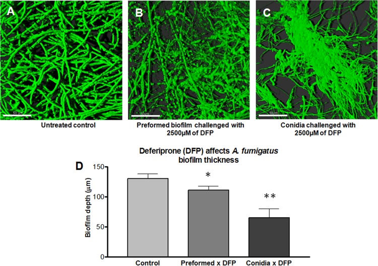
FIG 4.
Confocal laser scanning microscope (CLSM) images of A. fumigatus biofilm grown on polycarbonate disk surface. (A) Horizontal (xy) view of a reconstructed three-dimensional (3D) image of A. fumigatus biofilm (untreated control). (B) Horizontal (xy) view of a reconstructed 3D image of A. fumigatus preformed biofilm challenged with 2,500 μg/ml DFP. (C) Horizontal (xy) view of a reconstructed 3D image of A. fumigatus biofilm formation challenged with 2,500 μg/ml DFP. (D) Effect of DFP on A. fumigatus biofilm thickness. Assays were performed in triplicate, and images were taken from three different fields from each sample stained with FUN 1. The results are representative of two different experiments for each condition tested. The values shown are the means ± the standard deviations. The control, preformed × DFP, and conidia × DFP correspond to the conditions shown in panels A to C, respectively. One asterisk indicates a P value of <0.01, and two asterisks indicate a P value of <0.001 for the biofilm thickness compared to the untreated control. Magnification, ×63. Scale bar, 50 μm.

