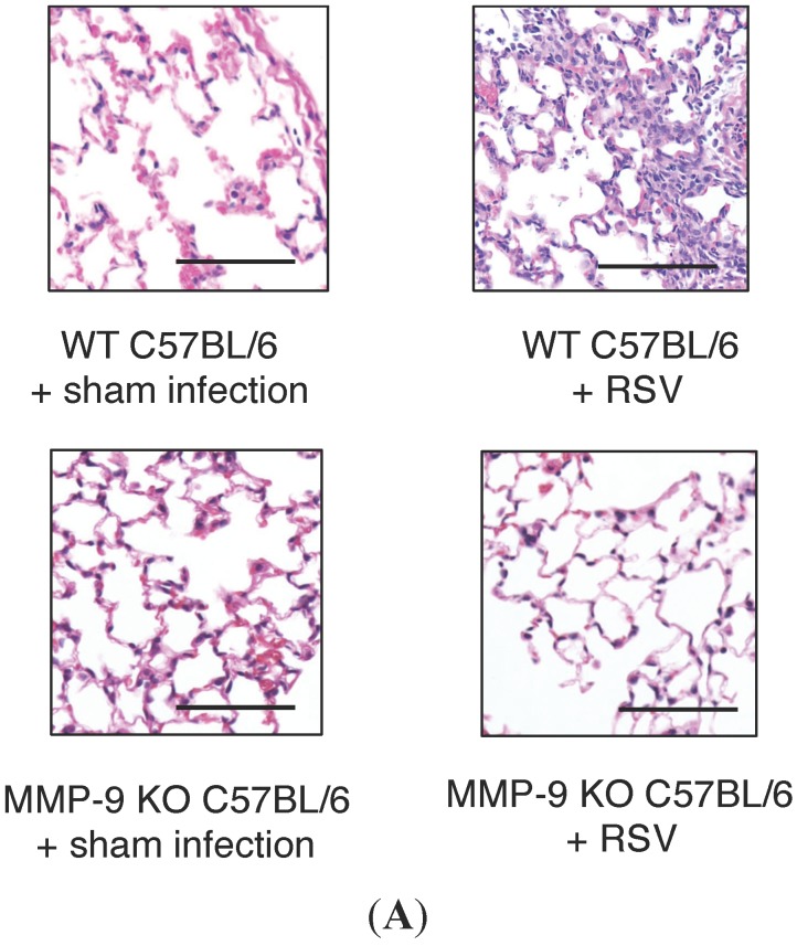
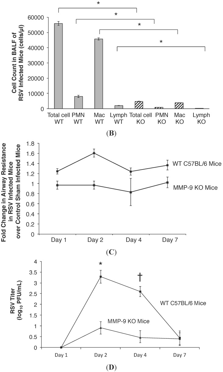
Figure 4.
(A) Lung inflammation in RSV infected WT and MMP-9 KO mice. WT and MMP-9 KO C57BL/6 mice received the same quantity of RSV inoculum. Tissue was stained with hematoxylin and eosin to assess lung inflammation post RSV infection. Acute pneumonitis with perivascular and peribronchial inflammatory infiltrates was observed in the RSV infected WT animals on days 2 PI (vs. normal lung histology in mock infected WT controls). In contrast, minimal lung inflammation was noted in the RSV infected MMP-9 KO mice. A similar histologic pattern was observed in RSV infected and sham mock infected MMP-9 KO mice. Scale bar: 67 μm; (B) Decreased cellularity in BALF following RSV infection in MMP-9 KO mice relative to WT mice. Total cell count in BALF of RSV infected MMP-9 KO mice (n = 5; stripe column) was almost 11-fold lower than RSV infected WT C57BL/6 mice (n = 5; grey column) on day 2 PI. Approximately 80% of cells found in both MMP-9 KO and WT mice were macrophages. The absolute macrophage count in the MMP-9 KO mice was 3814 vs. 45,737 cells/μL in the WT mice, respectively. In the MMP-9 KO mice, 19% of the total cell count was neutrophils (897 cells/μL), with lymphocytes accounting for less than 1% of cells recovered. In the WT mice, 14% of the total cell count was neutrophils (8060 cells/μL), and 4% was lymphocytes. Data represent mean ± SEM. * p < 0.04 as indicated, by ANOVA followed by Tukey-Kramer multiple comparisons test. PMN = neutrophils; Mac = macrophages; Lymph = lymphocytes; (C) Airway resistance is elevated in RSV infected WT mice compared to sham infected WT controls, but remains minimally changed following RSV infection in MMP-9 KO mice. WT and MMP-9 KO C57BL/6 mice (n = 10 per time point) were inoculated intranasally with107 PFU of live RSV, and airway resistance was measured from the time of inoculation to day 7 PI. Airway resistance was similar between MMP-9 KO mice inoculated with RSV vs. those given a sham infection throughout the duration of infection. This is in contrast to increased airway resistance measured in RSV infected WT mice over time of infection when compared to control sham infected mice. On day 1 PI, airway resistance was 1.24 fold higher in RSV infected WT mice compared to control mice. Peak airway resistance in the RSV WT mice was measured on day 2 PI, with a 1.6 fold increase in resistance measured compared to sham infected controls. Airway resistance remained increased on days 4 and 7 PI in the WT mice. Data represent mean ± SEM; (D) RSV titer in WT and MMP-9 KO mice following RSV infection. WT and MMP-9 KO C57BL/6 mice were inoculated with 107 PFU/mL of RSV, and viral titer was determined in BALF over multiple time points post infection. The RSV titer was approximately 2 logs lower in MMP-9 KO mice compared to WT mice on days 2 and 4 post infection. The viral burden was similar between MMP-9 KO and WT mice on day 1 and 7. Data represent mean ± SEM. * p < 0.001 and † p < 0.002 as indicated, by ANOVA followed by Tukey-Kramer multiple comparisons test.


