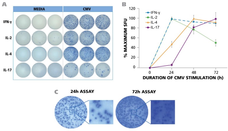
Figure 1.
PBMC from 45 donors was tested in ELISPOT assays without adding HCMV (antigen) or in the presence of UV-inactivated HCMV virions. The assay was performed as described in Materials and Methods with 250,000 PBMC per well and 50 μg/mL of HCMV antigen. (A) Representative well images of one donor for medium control and antigen-specific IFN-γ, IL-2, IL-4, and IL-17 assays are shown at the time point of peak secretion; (B) PBMC was stimulated with HCMV in ELISPOT assays for 24 h, 48 h, and 72 h. IFN-γ, IL-2, IL-4 and IL-17 responses exhibited as spot forming units (SFU) were recorded at each time point. (n = 3) (C) Maximal number of IFN-γ spots was observed at 24 h. At later time points (72 h) the ELISPOT image was over developed.

