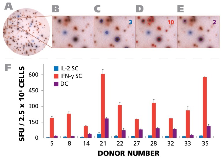
Figure 8.
Detection of polyfunctional CD4 cells. PBMC from 10 donors (IDs are specified in the figure) were subject to an IFN-γ/IL-2 Dual color ELISPOT assay at 250,000 cells per well, upon activation with 50 μg/mL of HCMV antigen. PBMC donor IDs were selected based on the response observed in Figure 4A. (A) A representative well image with both IFN-γ and IL-2 response is shown; (B) A digitally magnified portion of the representative well is shown; (C) Blue colored SFU representing IL-2 response are overlaid with a blue circle. The number of IL-2 SFU is inset in the figure; (D) Red colored SFU representing IFN-γ response are overlaid with a red circle. The number of IFN-γ response is inset in the figure; (E) Responses that are positive for both IL-2 and IFN-γ are shown as SFU that are both blue and red, and are visually represented by a purple color. The number of polyfunctional, i.e., positive for both, responses is inset in the figure; (F) The mean recall response for IFN-γ, IL-2, and polyfunctional responses for the individual donors and their respective SD from three representative wells is shown.

