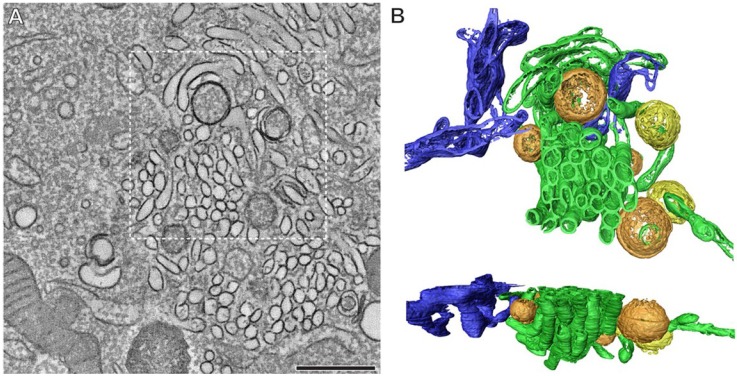
Figure 4.
Extensive membrane rearrangements in Enterovirus-infected cells. An electron tomographic slice through a serial tomogram, bar = 500 nm (A); and top and side views of the surface-rendered model of the boxed area (B) show the presence of single-membrane tubules (green), open (orange) and closed (yellow) double-membrane vesicles in a cell infected with coxsackievirus B3 at 5 h post infection. The ER is depicted in blue. Reprinted from Limpens et al. [70], mBio 2011 with permission from the authors, © 2011 by the American Society for Microbiology.

