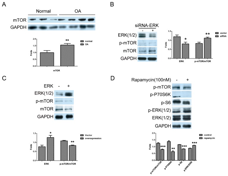
Figure 3.
The mutual inhibition of mTOR and ERK in human OA chondrocytes. (A) Normal and OA chondrocytes were cultured, and the level of mTOR was detected by western blotting analysis using anti-mTOR, p-mTOR, and GAPDH antibodies; (B) Cells were transfected with siRNA/ERK vector for seven days and the levels of mTOR and p-mTOR were detected by Western blotting analysis using anti-mTOR, p-mTOR and GAPDH antibodies; (C) Cells were transfected with ERK vector for two days and the levels of mTOR and p-mTOR were measured with western blotting analysis; (D) Cells were treated with rapamycin (100 nM) for 2 h and the levels of p-mTOR, p-P70S6K, p-S6, ERK(1/2), and p-ERK(1/2) were detected by western blotting analysis using anti-mTOR, p-mTOR, p-P70S6K, p-S6, ERK(1/2), p-ERK(1/2) and GAPDH antibodies. The blots were normalized to an endogenous protein (GAPDH). The values represent the mean ± SEM of three independent experiments (patients), each yielding similar results (* p < 0.05, ** p < 0.01, *** p < 0.001).

