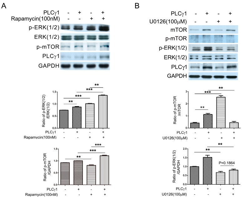
Figure 4.
The mutual inhibition of mTOR and ERK in PLCγ1-transformed human OA chondrocytes. (A) Cells were transfected with PLCγ1 vector for two days followed by the treatment of rapamycin (100 nM) for 2 h, and the levels of ERK(1/2), p-ERK(1/2), p-mTOR, and PLCγ1 were detected by western blotting analysis using anti-ERK(1/2), p-ERK(1/2), p-mTOR, PLCγ1 and GAPDH antibodies; (B) Cells were transfected with PLCγ1 vector for two days followed by the treatment of U0126 (100 μM) for 2 h and the levels of mTOR, p-mTOR, ERK(1/2), p-ERK(1/2) and PLCγ1 were detected by Western blotting analysis using anti-mTOR, p-mTOR, ERK(1/2), p-ERK(1/2), PLCγ1 and GAPDH antibodies. The blots were normalized to an endogenous protein (GAPDH). The values represent the mean ± SEM of three or five independent experiments (patients), each yielding similar results (** p <0.01, *** p < 0.001).

