
Figure 2.
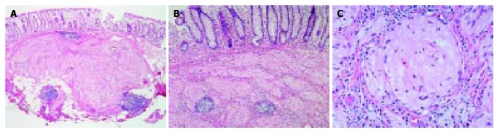
A submucosal tumor revealed by histological examination in resected tissue. A: Resected lesion showing a submucosal tumor covered with normal mucosa (HE staining, × 40). B: Higher magnification view of the tumor (HE staining, left: × 100; right: × 200).